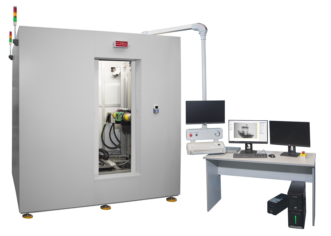

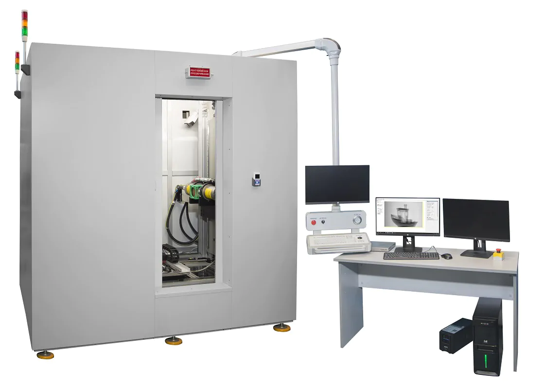

The “PIK-RKT-HR” full-size core X-ray tomograph is designed for high-resolution tomographic imaging of objects.
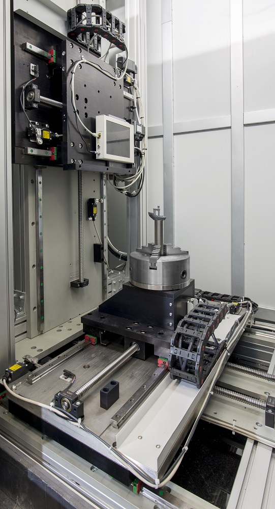
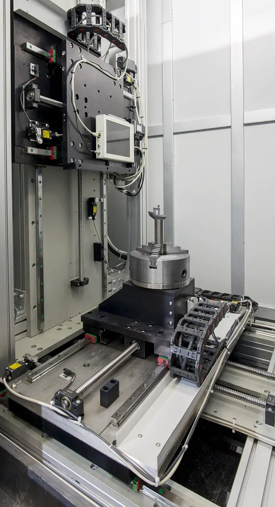
The tomograph is built on a scheme with independent movement of the source and detector, with servo drives used for all axes. The number of degrees of freedom is 7. The movement of the components is controlled using a joystick or via the control software. It also features passive protection for the sample movement drives, detectors, and tubes against mechanical damage.
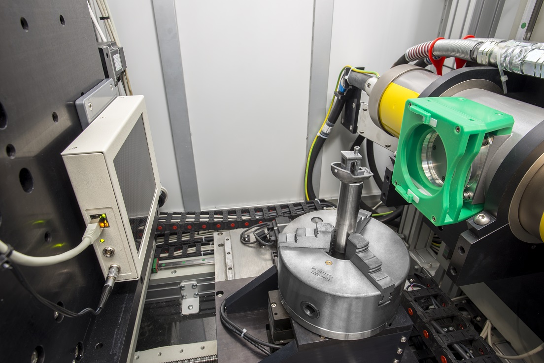
Structurally, the tomograph incorporates a passive anti-vibration system, including a granite base for the manipulators, detector, and X-ray tubes, as well as damping rubber supports. The core tube fixation system secures the object of study to achieve target characteristics for resolution and scanning speed.
The core analysis and core sample analysis software enables automated geometric calibration of the system in the selected axis position. It determines all parameters necessary for model reconstruction in that position with selected fixed geometric magnification, focal distance, and displacement of the object stage along the axis of rotation.
The results of the tomographic study of rocks include logging of fracture and vuggy porosity in the full-diameter core, assessment of the connectivity of fractures and vugs, determination of the spatial orientation of planar elements (fractures, bedding planes, etc.), and, in some cases, estimation of the mineral composition of the full-diameter core.
The full-diameter core analysis software allows for the visualization of selected density heterogeneities and their qualitative analysis. The tomography results (tomograms) are compatible with commercial spatial data analysis software products (such as Avizo).
The presence of a protective cabinet provides effective protection for personnel from the tomograph’s X-ray radiation and eliminates the need to place the device in a separate room. Measurements can be performed without extracting the core from the core barrel or the insulating tube.
Investigation of the three-dimensional (3D) structure of full-diameter core samples with a diameter of up to 120 mm in a protective tube with a diameter of up to 150 mm and a length of at least 1000 mm for:
Identifying and localizing discontinuities, fractures, vugs, man-made defects, the presence of bedding, and inclusions of “foreign bodies”.
Determining quantitative characteristics of fractures, such as thickness, length, aperture, geometry, spatial orientation, volumetric and linear density.
2D and 3D mapping of core sections and volumes, and delineating zones that are homogeneous in terms of X-ray absorption and structural-textural characteristics.
Investigation of rock samples with characteristic dimensions (in each direction) of 0.1–10 mm for:
Creating 3D digital models.
Assessing integrity.
Identifying and 3D mapping structural heterogeneities and micro-fracturing.
Determining capacity properties.
Creating digital models for the physico-mathematical simulation of filtration processes.
| General characteristics | |
| Positioning of the shooting object | vertical |
| Scanning | Vertical movement of the source-detector pair, rotation of the object |
| Maximum diameter of the object, mm | 200 |
| Maximum length of the object, mm | 1100 |
| Possible states of the core during shooting |
|
| X-ray source | |
| Number of simultaneously installed X-ray tubes | 2 |
| X-ray sources |
|
| Microfocus source | |
| Type | open type with unlimited service life with a two-stage air pumping system using a turbomolecular and oil-free forevacuum pumps, unipolar (with a grounded anode) |
| Tube voltage, kV | up to 225 |
| Tube power, W | up to 800 |
| Focal spot, µm | 6 |
| Sharp focus source | |
| Type | Metal-ceramic X-ray tube |
| Tube voltage, kV | up to 320 |
| Tube power, W | up to 1800 |
| Focal spot EN 12543, mm | 0,4 |
| Detector | |
| Type | high-contrast digital flat panel |
| Active area size, mm | 300×250 |
| Detector resolution, MP | 7 |
| Pixel size, µm | 99 |
| Capacity (grayscale), bits | 12 |
| Shooting speed, fps | 16-20 |
| Analog-to-digital converter (ADC) bit depth | 16 |
| Protective cabinet | |
| Design | Lead-steel sandwich panels, cellular structure of lead shielding, ensuring consistency of the thickness of the shielding sheets and consistency of shielding properties throughout the entire service life of the equipment. |
| Dose rate at any point at a distance of 100 mm from the cabinet surface, no more than, μSv/h | |
| Safety |
|
| Indication | Signal lights informing about the operation of the X-ray tube and located on the equipment body |
| Manipulator of the object under study | |
| Maximum travel along the axis, mm | 800 |
| Distance from the axis of rotation of the object to the X-ray tube (by the maximum dimensions of the X-ray tube), mm | from 0 to 400 |
| Distance from the axis of rotation of the object to the detector (by the maximum dimensions of the detector), mm | from 0 to 400 |
| Displacement resolution along the axes, µm | 1 |
| Rotation resolution, min | 5.4 |
| Detector manipulator | |
| Maximum travel along axis 1, mm | 800 |
| Distance from detector to X-ray tube (by max. dimensions of detector and X-ray tube), mm | from 0 to 800 |
| Maximum vertical travel (from rotating table and above), mm | 1000 |
| Travel along axis D3 (from center), mm | 400 (±200 from the center) |
| Displacement resolution along axes D1, D2, D3, µm | 1 |
| X-ray source manipulator | |
| Maximum vertical stroke (from the rotating table and above), mm | 1000 |
| Displacement resolution along the P1 axis, µm | 1 |